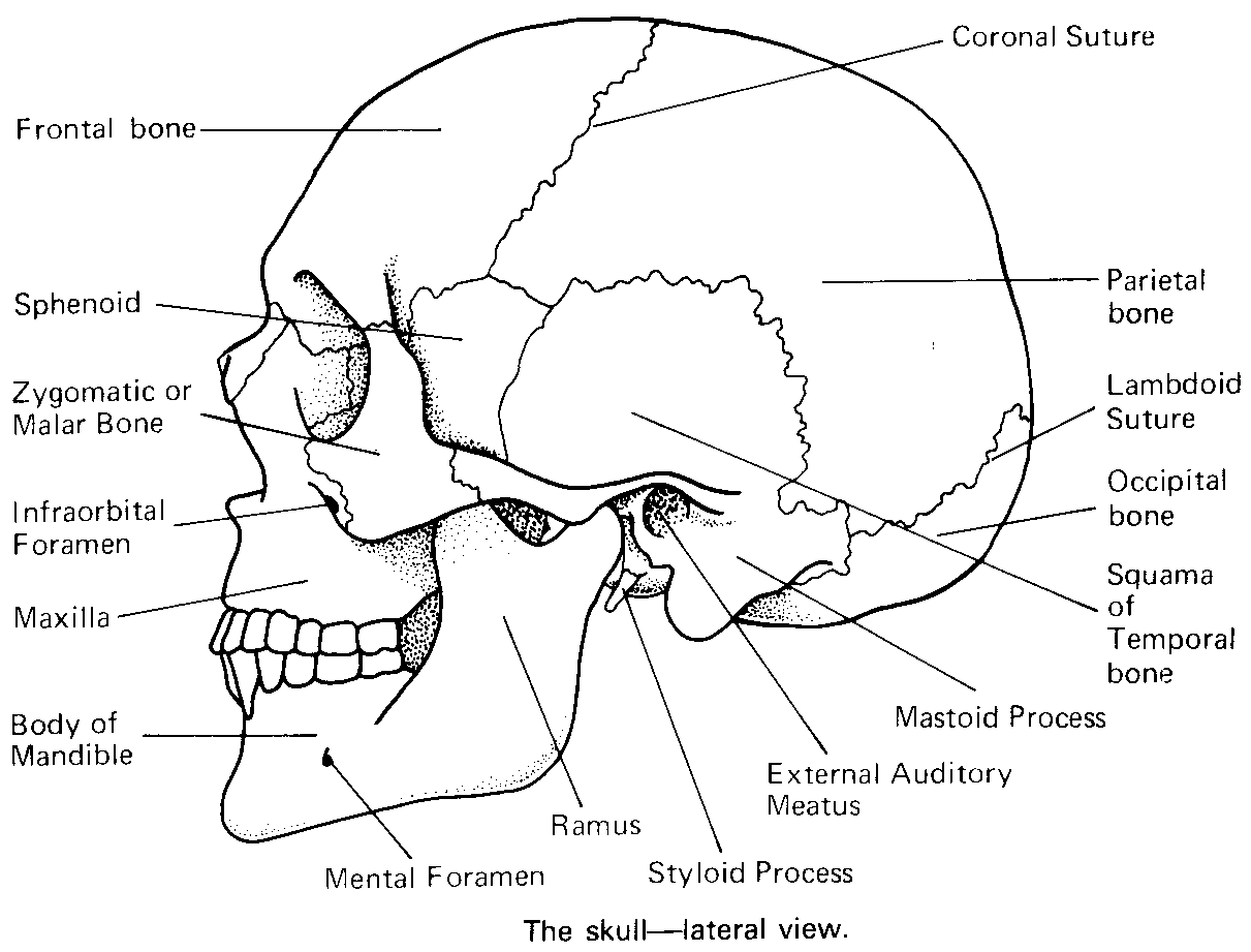
The skull is arguably the most complex area of our skeletal system. It is comprised of 22 bones–8 of which are cranial bones and 14 of which are facial bones. Together the cranial and facial bones protect and support our sensory organs, protect the brain, and provide a scaffolding for muscles. The intricate system of facial bones forms the mechanical framework that allows for the proper movement of the head and face.
The bones that make up the facial skeletal system include: the orbital bones (seven bones that enclose the eye), the nasal bones (two bones that form the bridge of the nose), the turbinate bone (spongy bones of the nasal cavity), the vomer (bone that separates the nasal passages), the zygoma (cheekbone, also known as the malar bone), the maxilla (upper jaw bone), and the mandible (the lower jaw bone).
Facial bones vary in size and strength. The mandible is the largest, sturdiest bone of the face, while the lacrimal bone, an orbital bone that supports the tear duct, is the smallest and most fragile. About the size of a fingernail, the lacrimal bone is, in fact, one the smallest bones in the body.

Fractures can occur on any bone of the face; however, the force it takes to break a facial bone depends on the bone’s strength as well as the speed and the angle of the impact. To help understand the nature of facial bone trauma, it may be helpful to consider the bones by region:
When compared to other bones of the body, nasal bones can be broken relatively easily. The most common facial bone trauma, nasal fractures account for up to 45% of all facial fractures.
Signs of nasal fractures: Swelling, tenderness, nasal deformity, bloody nose. In severe cases, the brain may be exposed to the outside environment.
Often the result of a blunt object hitting eye, orbital fractures can involve both the rim of the eye and the bone that keeps the eye in place. Damage to the eye socket is frequently seen in sports injuries.
Signs of orbital fractures: Tissue swelling and bruising, double vision, sunken eye.
A cheekbone fracture, often referred to as a zygoma fracture, is commonly combined with an orbital fracture. This type of fracture affects the functioning of the jaw and the position of the cheekbone.
Signs of cheekbone fractures: Flattened of the cheek with facial asymmetry, altered sensation around the injured area.
A lower jaw fracture is one of the most distressing facial fractures. Recovery from a mandible fracture can be lengthy and uncomfortable. Jaw trauma may also result in tooth damage and persistent facial numbness. Most commonly this injury results in difficulty and pain when either opening or closing the jaw.
Signs of mandible fractures: Jaw tenderness and pain, inability to bring teeth together and/or chew.
Fractures can occur on any bone of the face; however, the force it takes to break a facial bone depends on the bone’s strength as well as the speed and the angle of the impact. To help understand the nature of facial bone trauma, it may be helpful to consider the bones by region:

Although any type of impact can cause a bone injury, car accidents, sports trauma, falls, and assaults are the most common causes of facial fractures. Nasal and orbital fractures are common among boxers, martial arts fighters, and those participating in contact sports. Severe midface injuries and jaw fractures are often due to automobile crashes.
Ready to start looking your best? We offer virtual and in-office consultations.
Before recommending a treatment, it is important to properly diagnose a facial bone fracture. Depending on the area of the face affected, a simple X-ray, a CT scan, or a combination of both may be used.
X-rays are often useful in the initial diagnosis of jaw, orbital, and cheekbone fractures; additionally, they may be used to assess focal injuries (as in the case of a nasal fracture). When more information is needed, a CT scan may be ordered.
A computed tomography (CT) scan shows the detailed anatomy of the facial bones in excellent detail allowing the injury to be evaluated with a greater degree of accuracy.

Facial bone fractures are usually diagnosed using a CT scan. If the bones are found to be significantly out of position then surgery is usually recommended.
Injuries to the face or head often result following motor vehicle accidents, falls, assaults or sports-related trauma. If the force of the trauma is severe enough, facial bones can be broken. Broken bones in the face have the potential to impair a patient’s vision and their ability to chew, speak or even breathe. In addition, facial bone fractures can lead to obvious deformities that can cause significant problems if they are not repaired.
Facial bone fractures are usually diagnosed using a CT scan. If the bones are found to be significantly out of position then surgery is usually recommended. It is important that facial bone surgery only be performed by a surgeon with significant training in craniofacial and maxillofacial surgery. If facial bones are not repositioned properly, significant functional and aesthetic deformities can result.

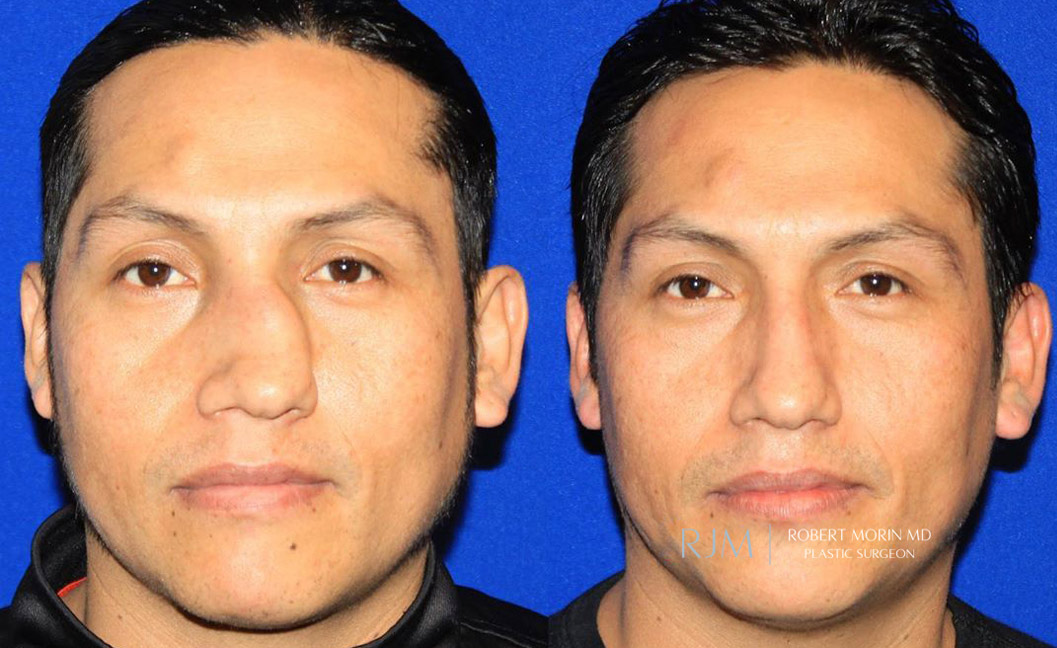
*Results may vary
Just as the facial bones vary, so does the treatment of facial bone fractures. Depending on the nature of the injury, extensive surgical repair may be required. This often involves the placement of titanium plates and screws in order to stabilize the bone. A craniofacial surgeon typically performs surgery on facial bone fractures. Using state-of-the-art technology, surgeon Dr. Robert Morin can comprehensively assess a facial bone injury and determine the appropriate surgical intervention.
Ready to start looking your best? We offer virtual and in-office consultations.