Discover giant congenital melanocytic nevus (GCMN ) treatment in New Jersey through innovative, ongoing, and safe surgical interventions performed by award-winning pediatric plastic surgeon, Dr. Morin.
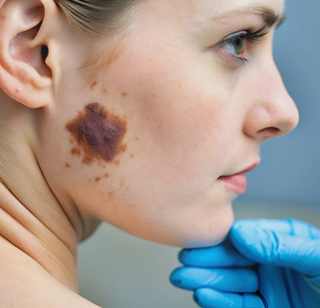
Giant congenital melanocytic nevi are large, pigmented, disfiguring skin lesions that are present at birth and that have a significant risk of developing malignant melanoma. In addition, these lesions result in significant psychological distress for both the patient and their family. These lesions occur at a rate of approximately one in 20,000 births and can involve the trunk, extremities, head, neck, and face. While these lesions are always present at birth, the extent of the lesions may not be initially appreciated due to changes in size, shape, and pigmentation over time.
Giant congenital nevus often appears as dark brown to black patches of skin covering the truck, neck, back, limbs, or face. Patches typically contain a defined, singular border whose surface area has hair and is markedly darker from its surrounding skin tone, making the contrast between the skin lesions and regular skin extremely pronounced.
It’s important to note giant congenital nevus patches will grow with the body. Children born with this condition often have proportional patches visible immediately or within one month of birth, with patches increasing in size as a child grows.
While patch sizes range, the condition is typically diagnosed early when a congenital nevus patch is around 10 centimeters. If untreated, lesions will continue to grow and darken, reaching up to 40 centimeters.
Giant congenital melanocytic nevus is considered a rare skin condition (1 in every 20,000 people) caused by gene mutations that occur at random during fetal development. This means giant congenital melanocytic nevus is non-hereditary — i.e. is not passed down between generations — but rather occurs randomly in-utero, called a somatic mutation.
Most cases are triggered by mutations within two specific genes, the NRAS and the BRAF. These genes are linked to producing melanocytes, the cells that create melanin and therefore your resulting skin tone. Current research indicates giant congenital melanocytic nevus is a result of overactive NRAS and BRAF genes overproducing melanocytes, though our understanding of this condition continues to evolve.
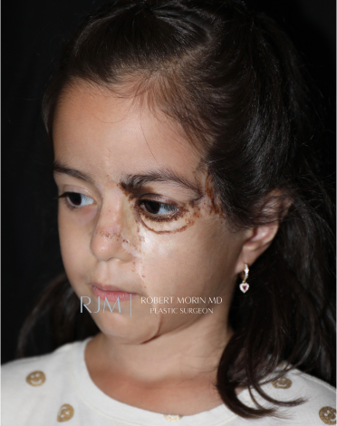
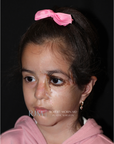
These before-and-after photographs showcase the patient’s transformation following surgical treatment performed entirely by Dr. Robert Morin. The results demonstrate the successful near-complete excision of the giant congenital nevus from the patient’s face, achieving an aesthetically pleasing reconstruction.
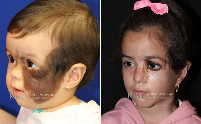
*Results may vary
The risk of developing melanoma skin cancer in these lesions over time has been recorded to be in the range of 2.8 to 8.5%.
This is a significantly higher risk than the risk in the general population and it is the main reason why complete surgical excision is generally recommended. The second reason why complete surgical excision is recommended is related to the severe psychological distress associated with these large pigmented skin lesions, especially those that involve the face.
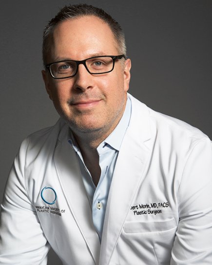
Dr. Morin is an American Board of Plastic Surgery board-certified plastic surgeon with specialized training in pediatric and craniofacial surgery, making him a clear choice for GCMN’s specific treatment approach in and around Hackensack, NJ.
Known for his precision and aesthetic focus, Dr. Morin delivers exceptional results for complex reconstructive cases, including congenital nevus removal, combining his advanced surgical expertise with compassionate pediatric clinical care during this skin condition’s important — and ongoing — surgical treatment timeline.

The goals of surgical excision are to decrease the risk of malignant melanoma, improve the aesthetic appearance of the patient, and maintain psycho-social well-being. Treatment modalities include serial excision where smaller portions of a large lesion are excised and directly closed over the course of several surgeries performed over time.
Tissue expansion is another extremely effective way of using extra skin to reconstruct areas where the skin has been excised. Skin grafting is also an effective way of replacing giant congenital melanocytic nevi with normal-appearing skin.
Many minimally invasive techniques have been described, however, these techniques leave cells behind and obscure the ability to monitor the patient for the development of skin cancer. As a result, these techniques are generally not recommended.
Regardless of the type of reconstruction performed, the goal of surgery in children with giant congenital melanocytic nevi is to have the surgical treatment completed by the time the child begins school.
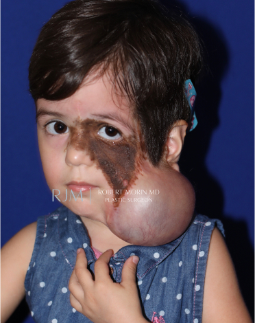
The patient seen below is one of Dr. Morin’s patients with a giant congenital nevus involving her face and scalp. The patient underwent surgical procedures over the course of several years in order to excise the giant congenital nevus from her face and to reconstruct her face using a combination of serial excision, tissue expansion, and skin grafting.
The photographs below demonstrate in detail the patient’s surgical journey from the initial diagnosis to school age. Surgery was begun early in this patient’s life in order to decrease her risk of melanoma and also to provide an aesthetically pleasing reconstruction by the time the patient entered school.
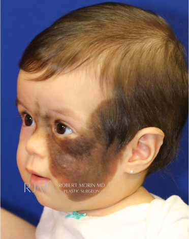
Here you can see the patient at a very young age during her initial consultation. At that time, a giant congenital melanocytic nevus involving the face and scalp was diagnosed and a surgical plan was discussed with the patient’s parents.
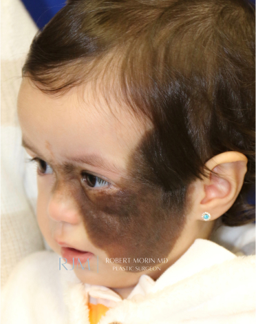
In this image you can see how these lesions evolve over time when the patient is young, both increasing in size and darkening in pigmentation.
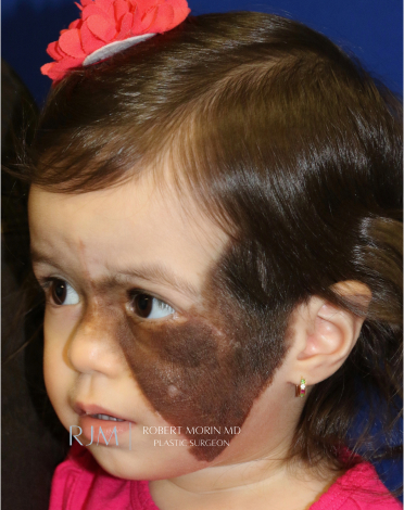
This photograph shows the patient after an initial serial excision was performed. The borders of the lesion were excised in order to create the ideal shape in preparation for tissue expansion.
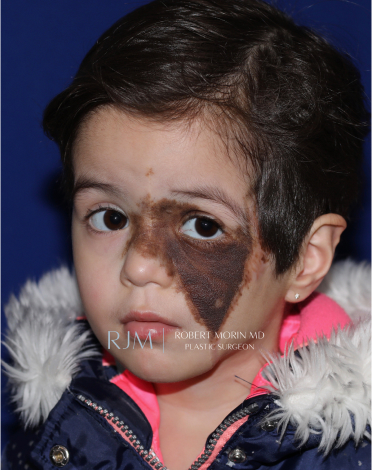
Here you can see the patient prior to her tissue expansion after several serial excisions had been performed.
The tissue expander is filled with normal saline over the course of several months to expand the overlying skin.
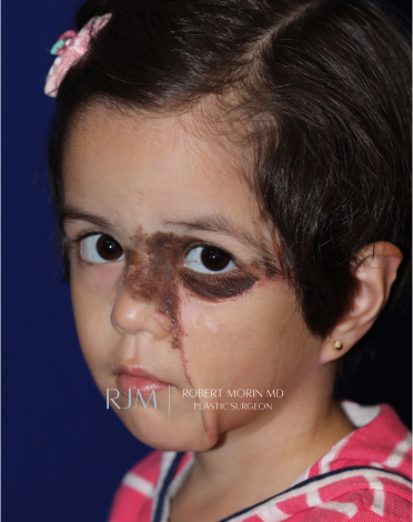
This is the result following the removal of the tissue expander and the reconstruction of her left cheek using the expanded skin.
In this photograph, you see the patient after additional excisions and full-thickness skin grafting to the left lower eyelid and nose.
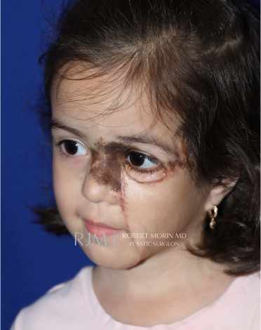
This is a photograph of the patient after more excisions and additional full-thickness skin grafting involving the nose.
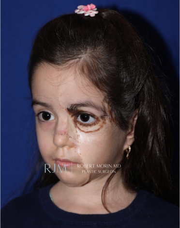
Here you can see the patient’s result after additional excisions and full-thickness skin grafting to the nose.
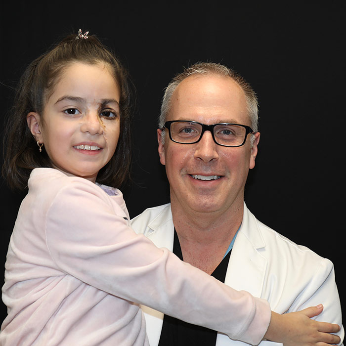
Final Excision and Full-Thickness Skin Grafting Results
Congenital nevus removal will typically require multiple surgeries staged over the years. While all surgeries carry some risks, these are reduced under Dr. Morin’s expert care and thorough patient-centric approach.
Dr. Morin and the pediatric plastic surgery team at RJM are here to support you across all physical, emotional, and psychological impacts of combating giant congenital nevus.
Giant congenital melanocytic nevus left untreated presents several risks and potentially serious side effects. These risks alter as a patient ages.
Dr. Morin and the team at Robert Morin MD offer personalized aftercare plans and ongoing support for patients undergoing Giant Congenital Melanocytic Nevus Treatment. This individualized post-surgical care addresses the following:
Clean surgical sites daily with antiseptic solutions and apply prescribed ointments to reduce infection risk and promote healing.
Administer pain relief medications as directed, especially to keep children comfortable during recovery.
Watch for redness, swelling, or unusual discharge, which may indicate infection or poor graft integration.
Keep grafted or expanded skin hydrated with physician-approved moisturizers to support elasticity and healing.
Limit strenuous activities to prevent tension on sutures or graft sites, ensuring proper healing.
Provide reassurance and age-appropriate explanations to help children cope with the physical and emotional demands of surgical recovery.
The risk of malignant transformation has been determined to be greatest in early childhood. For that reason, surgical excision has been recommended in children as young as six months of life. Contact Dr. Morin’s office for more information on ideal treatment options for giant congenital nevus.
No, melanocytic nevus do not go away on their own. Changes to nevi may occur with age, including changes to size and pigment. But the nevi itself is a near-permanent fixture of the skin requiring patient and doctor observation over time, particularly to flag melanoma or other skin cancer development signs.
A “nevus” is the most common medical term for moles. It’s important to note moles are extremely common, are found across the body, and are typically benign.
Common signs that a nevus or a patch of nevi may require medical attention include:
Note that complex skin conditions such as giant congenital nevus should be diagnosed and treated as early as possible.
The vast majority of GCMN patients live regular healthy lives, though it’s important to receive treatment for GCMN as early as possible. Early treatment, including surgical intervention to remove skin lesions, helps reduce your risk for developing melanoma and other aggressive forms of skin cancer, which GCMN patients are at a higher risk for. Surgeries can also help reduce additional cancer-type risks as well as lessen potential skin complications from GCMN lesions as you age.
Ready to start looking your best? We offer virtual and in-office consultations.